What is anal sac adenocarcinoma?
Anal sac adenocarcinoma is a malignant tumour that develops in the anal glands. This condition is more common in middle-aged to older dogs and often affects breeds such as cocker spaniels, beagles, and German shepherds. If not treated, the tumour can spread (metastasize) to local lymph nodes and other parts of the body.
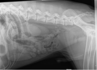
Why is a CT scan important?
A CT scan plays a crucial role in the prognostication of anal sac adenocarcinoma. It allows for a detailed assessment of the tumour size, regional lymph nodes, and any distant metastasis. Enlarged lymph nodes, especially the iliac lymph nodes, can be identified more readily on CT compared to ultrasound, providing important information for staging the cancer.
The CT scan helps guide the surgical plan by determining whether lymph node involvement or distant metastasis is present. The CT facilitates less invasive surgery as a map is then available to remove the mass and identify affected lymph nodes. This is critical because the presence of enlarged lymph nodes or metastasis can affect the prognosis and guide treatment decisions, such as whether additional surgery or chemotherapy is needed.
What is an anal sacculectomy?
An anal sacculectomy is a surgical procedure where both anal sacs are removed. This is the primary treatment for anal sac adenocarcinoma. Before surgery, the area around the anus will be thoroughly cleaned and clipped to prepare for the procedure (including the hair above the tail base). This is necessary to ensure that the surgery site is sterile, minimizing the risk of infection. An epidural will be administered to provide pain relief and muscle relaxation in the pelvic region. This ensures that your pet remains comfortable during the procedure, as the anal area will be the primary surgical site. The surgeon will use careful techniques to minimize trauma to surrounding structures, ensuring the best chance for a successful recovery.
Why is this surgery performed?
Anal sac adenocarcinoma is an aggressive tumour that can spread to nearby lymph nodes and other organs if not removed. Early surgical intervention is key to improving prognosis and managing symptoms. By removing both anal sacs and affected tissue, we aim to prevent further complications such as lymph node involvement and metastasis.
Prognostic information:
Clinical staging and prognosis
The TNM clinical staging system is used to assess tumour size and spread. This system takes into account the primary tumour size, regional lymph nodes, and distant metastasis. The stage allows us to estimate a median survival time (MST). This the time at which half of the patients with the disease are still alive. For example, if a dog with anal sac adenocarcinoma is given an MST of 24 months, this means that 50% of dogs in this group will live beyond 24 months, while the other 50% will have passed away by that time.
- Stage 1 & 2: these stages are based on the size of the primary tumour, with no evidence of metastasis. In stage 1, the tumour is smaller than 2.5 cm. In stage 2, the tumour is greater than 2.5 cm.
MST for dogs in stage 1 is 40 months, and for stage 2 it is 24 months. - Stage 3 & 4: in these stages, the primary tumour's size is irrelevant. The main difference between these stages is the involvement of regional lymph nodes and distant metastases:
- Stage 3a: regional lymph node metastasis is present but less than 4.5 cm in diameter.
MST for stage 3a is around 15-16 months. - Stage 3b: regional lymph nodes are larger than 4.5 cm in diameter.
MST for stage 3b is around 10-11 months. - Stage 4: distant metastasis is present, and the prognosis becomes more guarded. Median survival for this stage is typically lower and can be influenced by how well the metastasis is managed.
Chemotherapy
Chemotherapy is commonly used in combination with surgery to reduce the risk of recurrence, especially in cases where the tumour has spread to nearby lymph nodes or other areas. The goal of chemotherapy is to target any remaining cancer cells that surgery could not remove. Surgery is the mainstay treatment with the most evidence to elongate good quality life. Following surgery, we will refer you to an oncologist.
What to expect after surgery:
- Hospitalisation: your pet will stay overnight, to ensure pain management, monitor their initial recovery, and address any post-surgical concerns with our 24/7 on-site team.
- Recovery period: your dog will require a 2–4-week recovery period post-surgery. During this time, it’s important to limit activity to avoid strain on the surgical site. Given disruption of the anal sphincter faecal accidents are expected in the short term. This is not a long-term concern.
- Wound care: the incision site will need to be monitored for signs of infection, including swelling, redness, or discharge. The area may need to be gently cleaned.
- Diet: we advise the addition of metamucil (1-2 teaspoons over food) to soften stools and facilitate faecal passage.


.svg)
.svg)